Where science and art openly hangout

In this post:
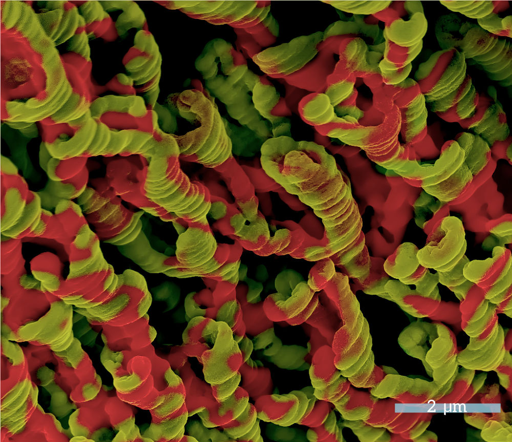
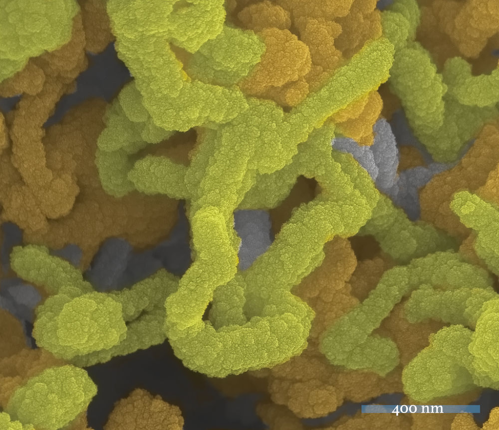
Some Scanning Electron Micrographs (SEMs) from those bygone days in which I would loose myself into the tiny world that only electrons unaided could see. Isn’t it weird to be nostalgic about metallic and semiconducting electrodes?
SEM stands for Scanning Electron Microscopy which is an imaging technique that uses electrons to obtain images of structures that are two small to be observed with visible light. People also call SEM the micrographs thus acquired. Basically, you bombard your material of interest with a beam of electrons of a certain energy and depending on the topography and composition of your target, these electrons interact with it in various ways that are then detected and translated into a visual representation of it. This is obviously, a simplified description of the imaging process; for in depth information there are many good sources online you can look up.
Anyway, I spent a good chunk of my professional life looking at various computers screens into the wonderful world of metallic and semiconducting electrodes, and trying to figure out how the different tiny structures looked and how to establish systematic relationships between the way that I prepared them, their morphology and chemical composition, and they way they performed in a couple of different devices. I obtained hundreds of images from a variety of materials, I harvested electrodes from rechargeable batteries cycled under different conditions, published a few papers. It is interesting how many images one captures before being able to glean some understanding of the processes under study, but at the end you publish only the most illustrative of them, a very small portion to be sure.
SEMs, because they are not produced with visible light, come in greyscale micrographs; you can add color electronically either with a modern instrument with software which would do that for you, or you can use a commercial image processing software to do it by yourself. The latter is what I have done with the two images in the post, they come from some work I did during my PhD more than twelve years ago. In their current format, these micrographs are mostly of artistic interest because isolated from the rest of the tests and analysis conducted on the sample, they don’t say so much, and also because we scientists don’t like things processed in ways in which we cannot do it ourselves if we ever wanted to.
The experiments can be replicated, new electrodes grown, and new images taken, but this color application is purely artistic and purely mine. Isn’t it beautiful? Enjoy!

Did you like this post?
Then you will probably like: